Modern oral healthcare relies heavily on advanced diagnostic technologies to detect underlying pathologies that cannot be identified through visual clinical inspection alone. As a foundational diagnostic tool in contemporary dentistry, a professional dental x-ray allows clinicians to evaluate the internal anatomy of teeth, root canal systems, periodontal ligament spaces, and alveolar bone architecture with exceptional precision. While routine visual examinations reveal surface enamel discolorations, obvious cavities, and superficial plaque accumulation, they provide no information regarding what occurs beneath the gumline or inside dense calcified structures. By capturing high-resolution radiographs of the oral cavity, a diagnostic dental x-ray helps dental professionals identify hidden interproximal decay, periapical abscesses, impacted wisdom teeth, bone resorption patterns, and developmental dental anomalies before they progress into debilitating dental emergencies. Incorporating these radiographic examinations into routine preventive visits ensures that minor dental issues are resolved conservatively, minimizing discomfort, treatment duration, and long-term healthcare expenses for patients of all ages.
What Is a Dental X-Ray and How Does Radiographic Imaging Work?
A radiographic examination in dentistry functions by directing a controlled beam of low-dose ionizing electromagnetic radiation through oral tissues onto an electronic digital sensor or traditional photographic film. As the x-ray photons pass through the mouth, different anatomical structures absorb the radiation at varying rates depending on their physical density. Dense calcified tissues, such as tooth enamel, dentin, and alveolar cortical bone, absorb the highest amount of radiation and appear light or radiopaque on the resulting image.
Conversely, soft tissues such as the dental pulp, gingiva, periodontal ligaments, and air spaces allow radiation to pass through unimpeded, causing them to appear dark or radiolucent on the display. Pathological conditions like dental caries, root infections, and bone loss demineralize hard tissues, creating darker radiolucent areas within normally bright white structures. This radiographic contrast provides clinicians with a clear anatomical map of the patient's dentition, allowing for accurate diagnostic analysis and tailored therapeutic interventions.
What Are the Primary Types of Dental X-Ray Procedures?
Bitewing Radiographs
Bitewing images focus specifically on the coronal portions of the upper and lower back teeth within a single view. They are primarily utilized during routine examinations to detect interproximal cavities forming between adjacent molars and to measure crestal bone levels associated with early periodontal disease.
Periapical Radiographs
Periapical projections capture the entire anatomical tooth structure from the biting crown to the root apex, including the surrounding alveolar jawbone. These images are essential for diagnosing root fractures, periapical abscesses, deep periodontal pockets, and endodontic pathoses requiring root canal treatment.
Occlusal Radiographs
Occlusal radiographs provide a broader perspective of the floor of the mouth or the hard palate. They track the development and positioning of entire arches of teeth, locate foreign objects or salivary gland stones, and identify supernumerary impacted teeth in growing pediatric patients.
How Do Extraoral Radiographs Differ From Intraoral Images?
While intraoral radiographs place the digital sensor inside the patient's oral cavity to capture close-up views of specific teeth, extraoral imaging systems keep both the radiation tube and the detector outside the mouth. Extraoral techniques provide broad anatomical surveys of the maxilla, mandible, temporomandibular joints, and facial skeletal framework. They are ideal for patients with severe gag reflexes, limited mouth opening capabilities, or extensive maxillofacial trauma.
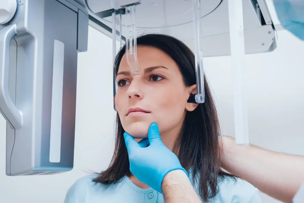
Panoramic radiographs, commonly known as orthopantomograms (OPG), wrap around the patient's head to deliver a comprehensive flat representation of the entire upper and lower jaw in a single frame. Cephalometric projections capture the entire lateral profile of the skull, documenting the spatial relationship between the jaws and the cranial base. These wide-angle extraoral imaging protocols are essential for planning orthodontic alignment, orthognathic surgical corrections, and complex surgical reconstructions.
Why Do Dentists Need a Dental X-Ray for Accurate Diagnosis?
- Detecting Interproximal Dental Decay: Identifying microscopic cavities developing between tightly contacting teeth where physical explorer probes and visual mirrors cannot reach.
- Evaluating Alveolar Bone Loss: Measuring the exact depth of crestal bone resorption caused by chronic periodontal infections to determine appropriate gum therapies.
- Identifying Root Canal Infections: Locating silent periapical radiolucencies and cyst formations at the root tip before painful acute facial swellings occur.
- Assessing Unerupted and Impacted Teeth: Mapping the precise three-dimensional angulation of impacted third molars relative to the inferior alveolar nerve canal prior to planned tooth extraction procedures.
- Monitoring Pediatric Dental Development: Checking for congenital tooth agenesis, assessing permanent tooth eruption paths, and identifying spatial crowding early.
Is Radiation Exposure From a Dental X-Ray Safe for Patients?
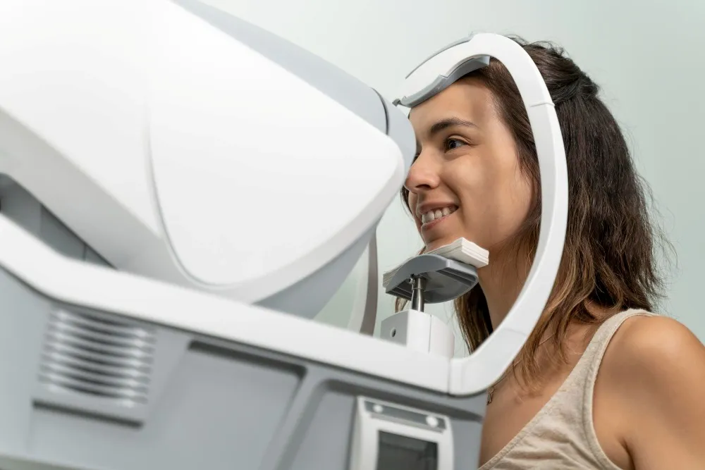
Concerns regarding radiation exposure in healthcare settings are common among patients, but modern dental radiography is among the safest diagnostic tools in clinical medicine. Radiation doses are measured in microsieverts, a scientific unit that quantifies biological exposure risk. A standard digital bitewing dental x-ray series produces approximately five microsieverts of radiation, which is equivalent to the natural background radiation every human absorbs from cosmic rays, soil minerals, and atmospheric radon over a typical single day.
Furthermore, contemporary dental practices strictly adhere to the ALARA principle—an international safety protocol dictating that exposure must remain "As Low As Reasonably Achievable." The integration of solid-state digital sensors, rectangular collimators that narrow the radiation beam, and high-frequency DC generators has reduced patient radiation dosages by up to eighty percent compared to traditional film imaging. Consequently, the biological health risk associated with diagnostic dental x-rays is exceptionally negligible compared to the substantial medical dangers of unmanaged oral infections.
What Safety Protocols and Protective Equipment Are Used During Imaging?
Lead Aprons and Thyroid Collars
Patients are shielded with lead-impregnated or lead-equivalent protective aprons that drape over the torso, alongside specialized thyroid collars designed to protect radiation-sensitive neck glands from secondary scatter radiation.
Digital CMOS and Phosphor Plate Sensors
Modern digital sensors require a fraction of the radiation energy needed by old chemical films, converting photon signals into computer screen pixels almost instantaneously with minimal beam duration.
Collimation and Beam Alignment Devices
Rectangular collimator cones restrict the emitted x-ray beam precisely to the dimensions of the digital sensor, preventing unnecessary radiation exposure to surrounding facial tissues.
How Often Should You Have a Routine Dental X-Ray Taken?
The clinical frequency of radiographic examinations is determined individually based on patient age, caries risk level, periodontal stability, and personal dental history. The American Dental Association and the Food and Drug Administration advise against a one-size-fits-all approach. For healthy adult patients with robust oral hygiene, low sugar consumption, and no active cavities, a routine bitewing dental x-ray interval of twelve to twenty-four months is generally sufficient.
In contrast, individuals categorized as high-risk dental patients—such as those with xerostomia (chronic dry mouth), rampant enamel demineralization, deep periodontal pockets, or extensive restorative dental work—may require radiographic evaluations every six to twelve months. Pediatric patients whose dental arches are actively expanding and whose primary enamel is thinner often require periodic monitoring to catch rapid interproximal decay early. Dental professionals evaluate clinical risk factors before ordering radiographs, ensuring each exposure is medically justified.
Can Pregnant Women Safely Undergo a Dental X-Ray Examination?
Preventive dental care, diagnostic examinations, and routine cleanings are completely safe and strongly recommended during pregnancy. Surging levels of estrogen and progesterone during gestation cause exaggerated inflammatory responses in gingival tissues, commonly leading to pregnancy gingivitis and localized pyogenic granulomas. Leaving dental infections untreated poses significant health risks to both the mother and the developing fetus, as systemic bacteremia can elevate the risk of low birth weight and preterm labor.
The diagnostic radiation beam utilized during a routine dental x-ray is focused strictly on the head, jaw, and facial structures, avoiding the pelvic and abdominal regions. When combined with a protective lead apron equipped with a thyroid shield, fetal radiation exposure is virtually unmeasurable. While elective aesthetic procedures can be postponed until after delivery, diagnostic imaging required to evaluate acute toothaches, suspected root abscesses, or facial trauma should proceed without hesitation to safeguard maternal health.
How Does 3D CBCT Imaging Advance Diagnostic Capabilities?
Standard two-dimensional radiographs compress three-dimensional anatomical structures into flat planar images, which can sometimes result in superimposition and optical distortion. Cone Beam Computed Tomography (CBCT) addresses this limitation by rotating a pyramid-shaped radiation beam around the patient's head, acquiring hundreds of planar projections that specialized computer software compiles into an interactive, three-dimensional volume model.
This cross-sectional imaging reveals bone width, trabecular bone density, internal root canal branching, and the precise paths of vital anatomical landmarks like the mental nerve and maxillary sinus cavities. For patients planning comprehensive tooth replacement, pre-operative 3D tomography is essential for mapping jawbone density prior to receiving specialized dental implants Turkey treatments. Similarly, full-arch rehabilitations like the All-on-4 technique depend on 3D CBCT scans to calculate the exact angulation of posterior implants, maximizing contact with available bone without invasive grafting.
What Can a Dental X-Ray Reveal About Periodontal Disease and Bone Loss?
- Horizontal Crestal Bone Resorption: Revealing uniform, generalized reductions in alveolar ridge height across entire quadrants of teeth suffering from chronic periodontitis.
- Vertical Infrabony Defects: Visualizing deep, localized trough-like bone destructions that run parallel to specific tooth roots, requiring targeted regenerative bone grafting.
- Furcation Involvements: Identifying bone destruction occurring within the bifurcation or trifurcation spaces where multi-rooted molars split into individual root structures.
- Subgingival Calculus Deposits: Detecting dense, calcified bacterial tartar spurs clinging to root surfaces deep beneath inflamed gingival margins.
- Widened Periodontal Ligament Spaces: Documenting mobility and early biomechanical failure caused by excessive occlusal trauma and bite imbalances.
How Do Digital X-Rays Compare to Traditional Photographic Films?
The transition from traditional photographic film to digital radiography represents one of the most significant technological advances in modern dental medicine. Traditional film-based x-rays required physical processing in darkrooms using chemical developing solutions and fixers, creating toxic chemical waste and requiring up to ten minutes of development time. Furthermore, traditional film could easily be scratched, lost, or degraded by environmental exposure over time.
Digital radiography uses solid-state electronic sensors that capture x-ray photon energy and transmit the data directly to a computer monitor within seconds. Clinicians can magnify specific root apices, adjust contrast levels, colorize tissue densities, and measure millimeter distances on screen. This digital transition provides higher diagnostic precision while significantly reducing chairside appointment times. The table below outlines the core technical and practical differences between these two methodologies:
|
Diagnostic Feature |
Traditional Film Radiography |
Modern Digital Radiography |
Clinical Patient Advantage |
|
Radiation Exposure Dose |
Standard baseline exposure |
70% to 80% reduction |
Significantly safer biological profile |
|
Image Acquisition Time |
5 to 10 minutes (chemical processing) |
Instantaneous (3 to 5 seconds) |
Shorter dental appointments |
|
Image Manipulation Tools |
None (static physical film) |
Magnification, contrast, inverted filters |
Early detection of microscopic pathology |
|
Ecological Environmental Impact |
Produces chemical waste and lead foils |
Completely chemical-free |
Eco-friendly healthcare model |
|
Archiving and Data Sharing |
Physical paper charts and sleeves |
Encrypted cloud-based digital files |
Seamless multi-specialist referrals |
How Does Pediatric Dental Radiography Protect Developing Smiles?
Children's oral cavities develop at a rapid pace, with primary teeth guiding the eruption pathways of developing permanent teeth beneath the gumline. Enamel layers on primary teeth are roughly half the thickness of adult permanent enamel, which means untreated interproximal decay can penetrate the inner dental pulp within a few months. Pediatric dental radiography allows clinicians to detect interproximal demineralization early, enabling remineralizing fluoride treatments before fillings become necessary.
Additionally, specialized pediatric radiographs monitor developmental milestones. They confirm the presence of all permanent tooth buds, check for congenitally missing incisors or premolars, and detect ectopic eruption paths that could damage adjacent roots. Dental teams utilize pediatric-sized digital sensors and tailor radiation exposures to match a child's smaller physical stature, ensuring safe, comfortable, and preventative pediatric care.
Can a Dental X-Ray Detect Oral Tumors, Cysts, and Systemic Pathologies?
Dentigerous Cysts and Odontogenic Keratocysts
Pathological fluid-filled cysts frequently develop silently around the crowns of unerupted wisdom teeth or canines. Left undetected, they can expand and hollow out large areas of the mandibular jawbone, increasing the risk of spontaneous fractures.
Ameloblastomas and Odontogenic Tumors
Benign but locally aggressive jaw tumors can expand within bone marrow spaces without causing pain or external swelling. A panoramic dental x-ray detects these lesions early as multi-locular radiolucent areas, facilitating conservative surgical enucleation.
Carotid Artery Calcifications and Atherosclerosis
Wide-view extraoral panoramic radiographs often capture the cervical soft tissue areas of the neck, revealing calcified atheroma plaques within the carotid bifurcation. Identifying these vascular calcifications early allows dentists to refer patients for cardiovascular evaluations, helping prevent strokes.
Why Do Endodontic Treatments Require Multiple Radiographs?
Root canal therapy requires high geometric accuracy because the internal pulp canal anatomy is complex, microscopically narrow, and completely invisible from the outside. Endodontic procedures rely on a series of specialized periapical radiographs taken throughout treatment to ensure optimal outcomes. An initial diagnostic radiograph confirms the diagnosis by visualizing the periapical bone inflammation, root curvature, and number of internal root canals.
During the procedure, working-length radiographs verify that endodontic files reach the exact apical constriction without perforating the root tip. After shaping and disinfection, a trial fit radiograph confirms that the gutta-percha filling material matches the canal length. Finally, a post-obturation radiograph documents that the entire root system is hermetically sealed against bacterial reinfection. This sequential radiographic approach ensures predictable, long-lasting tooth preservation.
What Signs and Symptoms Indicate You Urgently Need a Dental X-Ray?
- Throbbing, Spontaneous Dental Pain: Constant, deep-seated toothaches that worsen at night or when lying down, indicating pulpal inflammation or acute periapical infection.
- Prolonged Thermal Sensitivity: Sharp pain triggered by hot or cold food and drinks that lingers for more than ten seconds, suggesting deep caries near the pulp chamber.
- Swelling of the Gums or Facial Tissues: Localized gum boils (fistulas) draining pus or spreading facial cellulitis, which require immediate radiographic evaluation to locate the source of infection.
- Recent Maxillofacial Physical Trauma: Sports impacts, falls, or accidents that loosen teeth or cause suspected jaw fractures require radiographic examination to evaluate root stability.
- Severe Pain Behind the Molars: Radiating jaw pain, neck stiffness, and trismus (limited mouth opening) around third molar areas, signaling impacted wisdom tooth pericoronitis.
How Should Patients Prepare for an Upcoming Dental Radiograph?
Preparing for a dental radiographic examination is straightforward and requires no prior fasting or dietary adjustments. When you arrive for your appointment, inform your dental care team of any significant medical updates, recent surgeries, active medical conditions, or suspected pregnancy. You will be asked to temporarily remove metal objects from the head and neck area, including eyeglasses, removable partial dentures, tongue studs, nose rings, earrings, and metallic hairpins, as metallic items block radiation and produce white scatter artifacts that can obscure underlying structures.
During the scan, relax your facial muscles and focus on breathing gently through your nose to suppress any natural gag reflexes. The radiographer will position the digital sensor inside your mouth using an ergonomic alignment holder, then step behind a protective barrier to activate the exposure for a fraction of a second. The high-resolution radiographic images appear instantly on the chairside monitor, allowing your dentist to walk you through your anatomical structures, explain clinical findings, and design a personalized, preventive oral health plan.
Doctor Profile

Author and Medical Review
Dr. Melike KENTKURANOral and Maxillofacial Surgery Specialist
View ProfileThis content has been evaluated in accordance with Hospitadent Medical Publication Board standards for medical accuracy, currency and patient information.
Our Awards
See All ›2019 – 2020
Health Tourism Champion
Since its founding, Hospitadent has provided oral and dental care to patients travelling from every continent for health tourism. In the Service Exporters' Association research of 2019 and 2020, we ranked first in the Dental Health Services category. Within the same report we placed 15th among all healthcare institutions in 2019 and moved up to 10th in 2020. With uncompromising customer satisfaction, Hospitadent remains the healthcare institution that treats the highest number of international oral and dental patients.
2013
TEMOS International Quality Certificate
Guided by dedicated international departments, Hospitadent welcomes patients from abroad without any language barriers, offering assistance with accommodation, city and Bosphorus tours, concierge guidance, treatment logistics and medication support to keep satisfaction at its peak. With the goal of carrying the same quality standards to both Turkish and foreign patients, we received the TEMOS "International Quality in Dental Care" certificate in 2013—an accreditation granted by the organization that evaluates and certifies worldwide patient service quality based on the "Made in Germany" quality seal.
2020
Turquality Brand Support Program
For 16 years, the Turquality Brand Support Program has helped Turkish products build and promote their image abroad. Dental Group Hospitadent earned the right to join this initiative, taking an important step on its branding journey. Being the first dental healthcare group in a program that includes only five hospitals from the healthcare sector proves not only the quality image we project, but also the high standards we uphold.
2018
Stevie Awards – Most Innovative Customer Service
The Stevie Awards, recognized worldwide for honoring outstanding organizations and leaders, is the only international business awards program. In 2018, Hospitadent became the first hospital in Turkey to win a Stevie Award by receiving the "Most Innovative Customer Service of the Year" prize. Signature experiences such as Smiling Center, Digital Smile Design and Dental SPA played a decisive role in earning this accolade.
2019 – 2020
Service Exporters Association Award
Serving dental tourists from all over the world since its foundation, Hospitadent was honored by the Service Exporters' Association in the Champions of Service Exports program, which celebrates the rising value of Turkey's service industries. Thanks to its contribution to the national economy, Hospitadent received a certificate of achievement and ranked 15th among general healthcare institutions in 2019 and 10th in 2020, continuing to lead oral and dental treatments with an unwavering focus on patient satisfaction.
2021
Great Place To Work® Certified
Based on the employee experience and satisfaction survey completed by our teams, GPTW Turkey evaluated Hospitadent and awarded us the "Great Place To Work® Certified" distinction. As the first and only dental hospital holding this leading workplace culture certificate, Hospitadent continues its journey toward many more achievements.