Comprehensive diagnostic imaging serves as the cornerstone of modern oral healthcare, empowering clinicians to detect hidden pathologies, plan complex surgical interventions, and evaluate skeletal anatomy with precision. Among the most essential radiographic examinations used across contemporary dental clinics, a panoramic x-ray provides an expansive two-dimensional view of the entire oral cavity within a single visual capture. Capturing the maxillary and mandibular dental arches, temporomandibular joints, maxillary sinuses, and supporting alveolar bone structures, this extraoral diagnostic technique eliminates the discomfort of conventional intraoral sensors. Undergoing a periodic panoramic x-ray enables dentists to identify asymptomatic infections, monitor wisdom tooth impactions, evaluate bone density, and establish tailored preventive strategies for lifelong oral health.
What Is a Panoramic X-Ray and How Does Orthopantomography Work?
Clinically referred to as an orthopantomogram (OPG), this specialized imaging modality utilizes the fundamental principles of rotational panoramic tomography. During the brief scanning cycle, an X-ray generator and an electronic image receptor rotate simultaneously around the patient's head in opposite directions along a curved focal trough. The resulting panoramic x-ray displays a flattened, comprehensive image of the curved horseshoe-shaped anatomy of the human jaws.
Modern digital sensor technology translates X-ray photons directly into digital data files that appear instantly on high-definition computer monitors. By utilizing focal troughs aligned precisely with average human arch dimensions, the diagnostic panoramic x-ray brings the structures inside the focal zone into sharp clarity while blurring out unwanted anatomical structures situated in front of or behind the path of rotation.
What Are the Primary Indications for a Panoramic X-Ray in Modern Dentistry?
- Assessment of Impacted Third Molars: Visualizing the root curvature, depth, and spatial relationship of wisdom teeth relative to the inferior alveolar nerve canal.
- Pre-Surgical Implant Planning: Measuring vertical bone dimensions and assessing anatomical landmarks prior to placing dental implants in the jawbone.
- Detection of Osseous Pathologies: Identifying asymptomatic jaw cysts, odontogenic tumors, radiolucent periapical lesions, and bone metastases.
- Evaluation of Orthodontic Development: Tracking mixed dentition stages, missing tooth buds, supernumerary teeth, and ectopic eruption patterns in children.
- Diagnosis of Temporomandibular Joint (TMJ) Disorders: Evaluating condylar head symmetry, arthritic erosion, flattening, and structural discrepancies in the jaw joints.
Panoramic X-Ray vs Intraoral Periapical Radiographs: What Are the Key Differences?
Field of View and Anatomical Coverage
Intraoral periapical radiographs focus on two to three individual teeth, capturing coronal enamel, root morphology, and immediate surrounding periodontal ligament details with high localized spatial resolution. In contrast, a panoramic x-ray offers a broad perspective that includes the entirety of both upper and lower jaws, nasal fossae, and bilateral TMJs in one image.
Diagnostic Resolution for Interproximal Caries
Due to the lower spatial resolution and natural overlap of adjacent premolar contact points, a panoramic scan is not the primary diagnostic tool for discovering tiny enamel decay. Detecting early interproximal cavities or planning a conservative dental filling typically requires supplementary bitewing X-rays for microscopic enamel assessment.
Patient Comfort and Clinical Tolerance
Intraoral bitewings require placing rigid plastic sensor plates inside the mouth, which can trigger severe gag reflexes in sensitive patients or cause mucosal pain in individuals with small dental arches. The extraoral nature of a panoramic scan provides total comfort, making it an excellent diagnostic tool for children, elderly individuals, and patients with limited mouth opening.
What Is the Step-by-Step Procedure for Getting a Panoramic X-Ray?
The imaging workflow is straightforward, efficient, and completely non-invasive. The patient is asked to remove all metallic items from the neck and head region, including earrings, necklaces, glasses, hairpins, and removable dental appliances, as metallic objects generate radiopaque ghost artifacts on digital scans. The patient is fitted with a protective lead apron without a high thyroid collar to avoid beam obstruction.
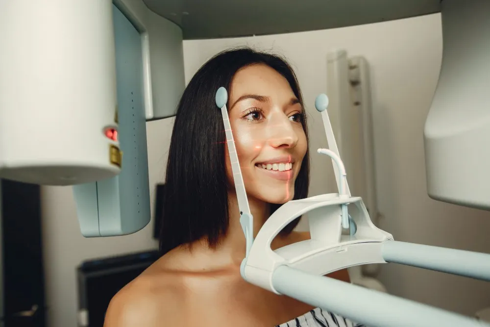
The patient stands or sits comfortably within the panoramic unit, placing their chin on a stabilized plastic rest and biting gently into a small sterile groove with their front incisors. The dental radiographer aligns the patient's head using vertical and horizontal laser positioning guides. The patient is instructed to press their tongue flat against the palate, close their lips, and remain motionless for approximately twelve to fifteen seconds while the machine completes its motorized rotation.
How Does Digital Dentistry Enhance the Quality of a Panoramic X-Ray?

The integration of advanced computer software into diagnostic radiology has transformed patient examinations. In the era of contemporary digital dentistry, traditional chemical developing tanks and darkrooms have been replaced by solid-state digital sensors that capture radiographs with remarkable clarity.
Digital imaging software allows clinicians to manipulate contrast, invert grayscale values, zoom in on suspicious bone lesions, and measure bone height down to fractions of a millimeter. Furthermore, digital storage allows instant transmission of radiographic records across specialized dental departments, making a digital panoramic x-ray an efficient component of comprehensive treatment planning.
Is a Panoramic X-Ray Safe and What Is the Radiation Dosage?
Dental radiographs utilize low ionizing radiation doses, adhering strictly to the ALARA principle (As Low As Reasonably Achievable). Modern digital panoramic units utilize narrow collimated X-ray beams and high-sensitivity sensors that reduce radiation exposure by up to 80% compared to traditional film-based radiography systems.
To put radiation exposure into clinical context, a standard digital panoramic scan delivers an effective radiation dose of approximately 10 to 24 microsieverts (µSv). This dosage is comparable to roughly two to three days of normal background radiation from our natural environment or equivalent to the radiation absorbed during a standard two-hour commercial airplane flight, confirming that a panoramic x-ray is safe for routine health screenings.
How Much Does a Panoramic X-Ray Cost Internationally?
The financial cost of diagnostic dental radiography varies depending on regional operational costs, clinical facilities, and whether the scan is billed independently or included within a comprehensive dental package. International dental centers in Turkey provide cost-effective diagnostic services, utilizing modern digital units at significantly lower fees than clinics in the UK, USA, or Australia.
|
Country |
Single Panoramic Scan (OPG) |
Full Diagnostic Checkup |
Standard Package Inclusions |
|
Turkey |
£25 – £45 / $30 – $60 |
£50 – £90 / $65 – $120 |
3D Panoramic Scan, Consultation & Plan |
|
United Kingdom |
£60 – £120 |
£130 – £250 |
Diagnostic Scan Only (Consultation Billed Extra) |
|
United States |
$100 – $250 |
$200 – $450 |
Image Only (Specialist Reading Fee Extra) |
|
Germany |
€65 – €130 |
€140 – €280 |
Standard Diagnostic Radiograph Fee |
|
Australia |
AU$90 – AU$180 |
AU$180 – AU$350 |
Diagnostic Imaging Only |
As outlined in the comparative table, undergoing diagnostic scans abroad offers significant value. Leading international clinics frequently include a digital panoramic x-ray within initial consultation packages to simplify treatment planning.
What Can a Panoramic X-Ray Reveal About Bone Health and Periodontitis?
Periodontal disease is a chronic inflammatory condition that leads to progressive destruction of the supporting alveolar bone. While clinical periodontal probing measures soft tissue pocket depths, a panoramic scan provides a visual overview of generalized horizontal and vertical bone loss patterns across both dental arches.
By comparing the height of the alveolar crest to the cementoenamel junction of each tooth, dentists can evaluate the overall severity of periodontal disease. A panoramic x-ray helps clinicians identify furcation involvements in multi-rooted molars, detect subgingival calculus deposits, and monitor bone stability following periodontal therapy.
How Does a Panoramic X-Ray Assist in Diagnosing Wisdom Teeth Problems?
Evaluating Impaction Angulation and Position
Wisdom teeth frequently lack sufficient arch length to erupt properly, becoming impacted in mesioangular, distoangular, horizontal, or vertical orientations. A panoramic scan displays the complete spatial position of third molars within the ascending ramus or maxillary tuberosity, aiding surgical extraction planning.
Mapping the Inferior Alveolar Nerve Canal
The primary clinical challenge during mandibular third molar surgery is avoiding neurological injury to the inferior alveolar nerve. A panoramic x-ray reveals critical signs of proximity, such as narrowing of the nerve canal, deflection of roots, or diversion of the canal path, indicating when 3D CBCT imaging is required.
Detecting Associated Follicular Cysts and Resorption
Impacted wisdom teeth can develop pericoronal pathologies, including dentigerous cysts, or cause external root resorption on adjacent second molars. A routine panoramic scan identifies these radiolucent follicular changes before they cause extensive bone destruction.
Can a Panoramic X-Ray Detect Temporomandibular Joint (TMJ) Disorders?
The temporomandibular joint is one of the most active joints in the human musculoskeletal system, susceptible to degenerative remodeling, inflammatory arthritis, and trauma. While soft tissue disc displacements are best visualized using Magnetic Resonance Imaging (MRI), a panoramic scan provides an excellent bony screening view of the bilateral condylar processes and mandibular fossae.
Clinicians evaluate condylar asymmetry, osteophyte formation, surface flattening, subchondral sclerosis, and structural hypoplasia. By providing simultaneous visualization of both the left and right joints, a panoramic x-ray assists in assessing condylar anatomy during jaw function.
What Are the Limitations of a Panoramic X-Ray in Diagnosis?
- Two-Dimensional Representation: Produces a 2D flattening of complex 3D skeletal structures, resulting in anatomical magnification and minor distortion.
- Lower Spatial Resolution: Lacks the fine pixel resolution required to detect early recurrent decay along dental margins.
- Superimposition of Ghost Images: Dense structures from the opposing jaw or spine can cast faint shadow artifacts across anterior teeth.
- Focal Trough Sensitivity: Poor patient positioning can cause anterior teeth to appear blurred, artificially narrow, or magnified.
- Limited Soft Tissue Detail: Cannot visualize the articular disc of the TMJ, mucosal tissue thickness, or individual muscle fibers.
How Frequently Should You Get a Panoramic Scan?
The recommended frequency for receiving a panoramic radiograph is personalized based on age, dental history, risk profile, and clinical symptoms rather than an arbitrary calendar schedule. For healthy adult patients with no active periodontal disease, low caries risk, and no recent oral surgeries, a routine panoramic scan is generally taken every three to five years to screen for silent osseous pathologies.
However, patients undergoing active orthodontic therapy, candidates planning dental implants, or individuals with chronic periodontal conditions may require more frequent imaging. The dental specialist determines when a panoramic x-ray is clinically indicated to minimize unnecessary exposure while ensuring diagnostic thoroughness.
Can Pregnant Women Undergo a Panoramic X-Ray Safely?
Elective diagnostic radiographs are generally postponed until after delivery to avoid unnecessary fetal exposure. However, when a dental emergency arises—such as acute facial trauma, severe spreading infection, or an agonizing impacted tooth abscess—diagnostic imaging becomes necessary to safely guide treatment.
Because the primary X-ray beam of a panoramic machine is directed exclusively at the maxillofacial structures, radiation scatter to the abdominal region is negligible. When paired with a lead apron with abdominal shielding, undergoing an emergency panoramic x-ray poses minimal risk to the developing fetus.
What Are Common Positioning Errors That Affect Image Quality?
Failure to Rest Tongue Against the Hard Palate
If the patient does not press their tongue against the roof of their mouth throughout the exposure, a dark radiolucent airway shadow is cast across the roots of the upper maxillary teeth, obscuring periapical anatomy.
Incorrect Frankfort Horizontal Plane Alignment
Positioning the patient's chin too low results in an exaggerated curved smile line with blurred mandibular incisors. Conversely, tilting the chin too high flattens the occlusal plane and can superimpose the hard palate over maxillary root tips.
Slumping or Incorrect Spinal Posture
When a patient slumps their shoulders instead of standing straight, the cervical spine absorbs X-ray photons, generating a wide radiopaque pyramid artifact across the midline that obscures the anterior teeth.
Why Do Modern Dental Clinics Rely on Panoramic Imaging?
Panoramic technology remains an indispensable diagnostic tool in contemporary dentistry, offering rapid imaging, low radiation dosage, and a wide diagnostic scope. It allows dental specialists to communicate findings clearly with patients, displaying full-mouth anatomy on screen during consultations.
From screening for asymptomatic bone pathologies to planning complex restorative rehabilitations, a digital panoramic x-ray provides an essential clinical overview. By laying the structural groundwork for comprehensive treatment planning, panoramic imaging supports predictable, safe, and effective dental care.
Doctor Profile

Author and Medical Review
Dr. Melike KENTKURANOral and Maxillofacial Surgery Specialist
View ProfileThis content has been evaluated in accordance with Hospitadent Medical Publication Board standards for medical accuracy, currency and patient information.
Our Awards
See All ›2019 – 2020
Health Tourism Champion
Since its founding, Hospitadent has provided oral and dental care to patients travelling from every continent for health tourism. In the Service Exporters' Association research of 2019 and 2020, we ranked first in the Dental Health Services category. Within the same report we placed 15th among all healthcare institutions in 2019 and moved up to 10th in 2020. With uncompromising customer satisfaction, Hospitadent remains the healthcare institution that treats the highest number of international oral and dental patients.
2013
TEMOS International Quality Certificate
Guided by dedicated international departments, Hospitadent welcomes patients from abroad without any language barriers, offering assistance with accommodation, city and Bosphorus tours, concierge guidance, treatment logistics and medication support to keep satisfaction at its peak. With the goal of carrying the same quality standards to both Turkish and foreign patients, we received the TEMOS "International Quality in Dental Care" certificate in 2013—an accreditation granted by the organization that evaluates and certifies worldwide patient service quality based on the "Made in Germany" quality seal.
2020
Turquality Brand Support Program
For 16 years, the Turquality Brand Support Program has helped Turkish products build and promote their image abroad. Dental Group Hospitadent earned the right to join this initiative, taking an important step on its branding journey. Being the first dental healthcare group in a program that includes only five hospitals from the healthcare sector proves not only the quality image we project, but also the high standards we uphold.
2018
Stevie Awards – Most Innovative Customer Service
The Stevie Awards, recognized worldwide for honoring outstanding organizations and leaders, is the only international business awards program. In 2018, Hospitadent became the first hospital in Turkey to win a Stevie Award by receiving the "Most Innovative Customer Service of the Year" prize. Signature experiences such as Smiling Center, Digital Smile Design and Dental SPA played a decisive role in earning this accolade.
2019 – 2020
Service Exporters Association Award
Serving dental tourists from all over the world since its foundation, Hospitadent was honored by the Service Exporters' Association in the Champions of Service Exports program, which celebrates the rising value of Turkey's service industries. Thanks to its contribution to the national economy, Hospitadent received a certificate of achievement and ranked 15th among general healthcare institutions in 2019 and 10th in 2020, continuing to lead oral and dental treatments with an unwavering focus on patient satisfaction.
2021
Great Place To Work® Certified
Based on the employee experience and satisfaction survey completed by our teams, GPTW Turkey evaluated Hospitadent and awarded us the "Great Place To Work® Certified" distinction. As the first and only dental hospital holding this leading workplace culture certificate, Hospitadent continues its journey toward many more achievements.