Understanding the differences between mucosal lesions is essential for effective pain relief and proper oral healthcare, which is why this complete guide to mouth ulcers (aphthous & traumatic ulcer): causes, symptoms & treatment breaks down everything you need to know about identification, management, and prevention. Oral mucosal ulcers are among the most common lesions encountered in clinical dental practice, affecting individuals of all ages across the globe. Whether triggered by an accidental bite during a meal, a sharp orthodontic wire, a nutritional deficiency, or an underlying systemic condition, these painful oral sores can significantly disrupt daily routines such as eating, drinking, speaking, and maintaining oral hygiene. While most mouth sores heal spontaneously without leaving permanent scars, persistent or recurrent ulcerations require a systematic diagnostic evaluation to rule out complex systemic diseases or malignant transformations.
By understanding the physiological mechanisms, environmental triggers, and clinical variations of both aphthous stomatitis and mechanical traumatic ulcers, patients and healthcare providers can better collaborate on targeting root causes rather than merely suppressing symptoms. This comprehensive reference explores the clinical characteristics of non-contagious canker sores and injury-induced mucosal lesions, highlights the role of daily oral hygiene products like SLS-containing toothpastes, outlines diagnostic criteria and coding standards, and offers evidence-based strategies for treatment and home care.
What Is an Aphthous Ulcer (Canker Sore)?
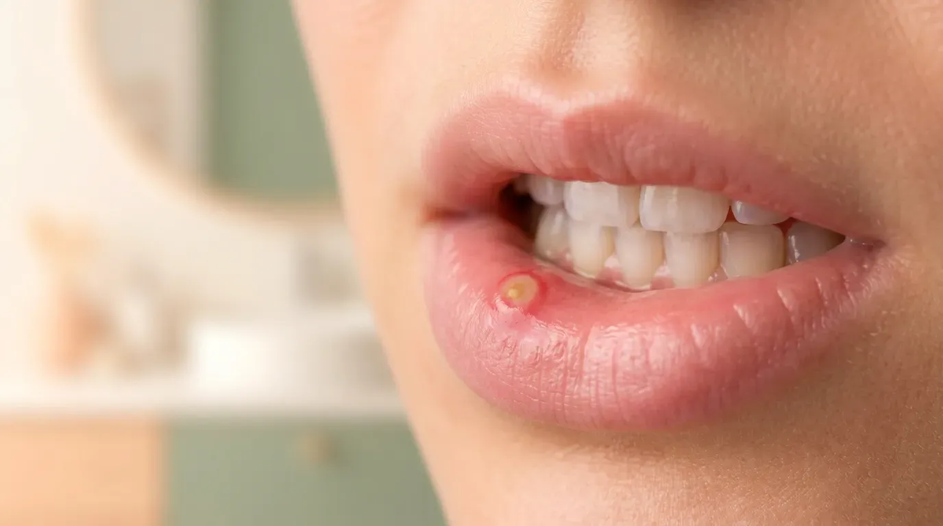
An aphthous ulcer, commonly referred to as a canker sore or recurrent aphthous stomatitis (RAS), is a painful, non-contagious inflammatory lesion that forms on the non-keratinized mucosal surfaces of the oral cavity. Unlike lesions associated with infectious pathogens, aphthous ulcers are mediated by the body's immune system, specifically involving T-cell-driven immune responses that result in localized tissue destruction. These ulcers characteristically present as round or oval craters with a necrotic center covered by a grayish-white or yellow pseudomembrane, surrounded by a distinct halo of bright red, inflamed tissue (erythematous border).
Clinically, aphthous stomatitis is classified into three primary variants based on size, depth, duration, and scarring potential:
- Minor Aphthous Ulcers: Accounting for approximately 80% to 85% of all cases, minor aphthae measure less than 10 millimeters in diameter. They are typically shallow, cause mild to moderate pain, and heal spontaneously within 7 to 14 days without leaving behind scar tissue.
- Major Aphthous Ulcers (Sutton's Disease): Representing about 10% of cases, major aphthous ulcers exceed 10 millimeters in diameter and penetrate much deeper into the submucosal tissue layers. These lesions are extremely painful, can persist for several weeks or even months, and frequently leave significant fibrous scarring upon resolution.
- Herpetiform Aphthous Ulcers: Making up 5% to 10% of cases, this subtype is characterized by clusters of numerous pinpoint ulcers (ranging from 1 to 3 millimeters) that may coalesce into larger, irregular ulcerated fields. Despite the name, herpetiform aphthae are not caused by the herpes simplex virus.
Aphthous Ulcer vs. Cold Sore (Herpes Simplex / HSV): Key Differences
|
Clinical Feature |
Aphthous Ulcer (Canker Sore) |
Cold Sore (Herpes Simplex / HSV-1) |
|
Etiology |
Non-contagious; immune-mediated inflammation |
Highly contagious; viral infection (HSV-1) |
|
Primary Location |
Non-keratinized mucosa (inner lips, cheeks, tongue, soft palate) |
Keratinized surfaces (vermilion border of lips, hard palate, outer skin) |
|
Initial Lesion |
Flat red spot progressing directly to an open ulcer |
Fluid-filled vesicles (blisters) that rupture and crust |
|
Contagiousness |
Not contagious; cannot spread to others |
Highly contagious through direct contact or fluid exposure |
|
Systemic Symptoms |
Rarely present in minor forms |
Prodromal tingling/burning, fever, localized lymphadenopathy |
|
Healing Time |
7–14 days (minor type); no scarring |
7–12 days; crusting stage followed by re-epithelialization |
|
Primary Treatment |
Anti-inflammatory agents, mucosal protectants, analgesics |
Antiviral medications (acyclovir, valacyclovir) |
One of the most widespread misconceptions in oral health is confounding aphthous ulcers (canker sores) with cold sores (fever blisters). Distinguishing between these two conditions is crucial because their underlying causes, contagiousness, and treatment approaches are entirely different.
Cold sores are caused by a contagious viral infection, primarily Herpes Simplex Virus Type 1 (HSV-1). After initial exposure, the virus remains dormant within the trigeminal ganglion and periodically reactivates in response to triggers like ultraviolet radiation, fever, stress, or immune suppression. Cold sores initially present as fluid-filled vesicles that rupture to form crusty lesions, located almost exclusively on keratinized epithelial surfaces such as the vermilion border of the lips, outer skin surrounding the mouth, hard palate, or attached gingiva.
In contrast, aphthous ulcers are non-infectious, non-contagious immune-mediated inflammatory reactions. They never form fluid-filled blisters prior to ulcerating, and they occur almost exclusively on non-keratinized, movable oral mucosa—such as the inner lips, labial mucosa, ventral tongue, floor of the mouth, soft palate, and buccal mucosa.
Causes of Canker Sores on the Tongue and Gums
The exact etiology of aphthous ulcers remains multifactorial, representing a complex interplay between genetic predisposition, environmental triggers, and immune dysregulation. When canker sores erupt on vulnerable non-keratinized tissue such as the ventral surface of the tongue or the unattached alveolar mucosa adjacent to the gums, several predisposing factors are frequently identified:
- Nutritional Deficiencies: Inadequate intake or poor absorption of essential micronutrients—particularly Vitamin B12, folate (Vitamin B9), iron, and zinc—can impair mucosal cell turnover and weaken the protective epithelial barrier of the mouth.
- Genetic Predisposition: Individuals with a family history of recurrent aphthous stomatitis have a significantly higher likelihood of developing lesions. Up to 40% of patients with frequent outbreaks report immediate family members affected by similar conditions.
- Psychological Stress and Anxiety: Elevated psychological stress stimulates cortisol secretion and alters mucosal immune surveillance, making the oral epithelium more susceptible to autoimmune-mediated localized destruction.
- Co-occurring Oral Inflammation: When soft tissues around the gums become chronically inflamed due to conditions such as gingivitis, micro-abrasions from brushing or chewing can more easily trigger localized ulceration on the compromised marginal mucosa.
- Hormonal Fluctuations: Many women experience recurrent aphthous outbreaks during specific phases of the menstrual cycle or luteal phase, correlated with progesterone drop-offs, while symptoms often improve during pregnancy.
- Dietary Hypersensitivities: Certain food groups containing high amounts of organic acids, nitrates, or specific proteins can trigger localized mucosal inflammation. Common culprits include citrus fruits, pineapples, tomatoes, walnuts, dark chocolate, and highly spiced foods.
- Microtrauma and Plaque Buildup: Minor, unperceived injuries to the delicate oral mucosa—such as minor abrasions from hard food crusts or calcified plaque build-up—can act as localized focal points for immune activation in susceptible individuals. Scheduling routine professional teeth cleaning and tartar treatment helps eliminate rough calculus deposits that irritate soft tissues.
What Is a Traumatic Ulcer and How Does It Form?
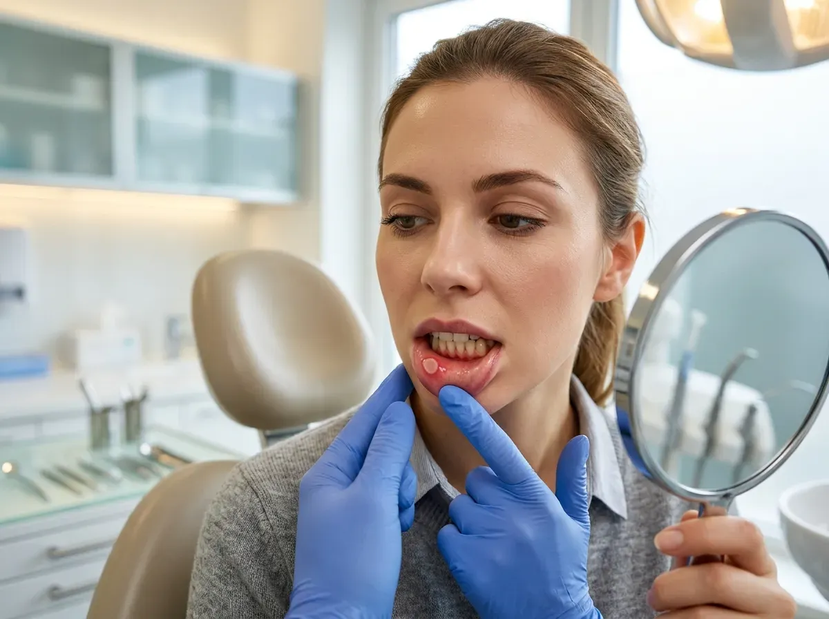
A traumatic ulcer is an oral mucosal injury resulting directly from physical, mechanical, thermal, or chemical damage to the protective epithelial surface. Unlike aphthous ulcers, which develop due to an internal immune mechanism without obvious external injury, traumatic ulcers have a clear, identifiable external cause. Once the surface epithelium is damaged or stripped away, the underlying vascular connective tissue (lamina propria) becomes exposed, initiating an inflammatory response that manifests as a painful ulcer.
Traumatic ulcers can occur on any anatomical surface of the oral mucosa, including both keratinized and non-keratinized tissues. Clinically, acute traumatic ulcers typically present as solitary, painful lesions with irregular, jagged, or linear borders corresponding to the shape of the offending agent. The ulcer bed is usually covered by a yellowish-white fibrin exudate, surrounded by variable degrees of erythematous inflammation. If the source of physical trauma is removed quickly, traumatic ulcers usually show rapid improvement and resolve completely within 7 to 10 days. However, if chronic physical irritation persists, the tissue may undergo chronic hyperplastic changes, developing raised, indurated (hardened) rolled edges that closely mimic the clinical appearance of squamous cell carcinoma.
Mouth Sores Caused by Biting, Braces, and Dentures
|
Characteristic |
Traumatic Ulcer |
Aphthous Ulcer (Canker Sore) |
|
Primary Trigger |
Known mechanical, thermal, or chemical injury |
Complex immune/genetic/systemic response |
|
Lesion Shape |
Irregular, jagged, linear, or matching irritant |
Symmetrical, round or oval crater |
|
Border Features |
Variable red border; may become rolled/hardened if chronic |
Sharp margin with a bright red halo |
|
Tissue Location |
Any oral site (tongue, palate, gingiva, cheeks) |
Almost exclusively non-keratinized mucosa |
|
Number of Lesions |
Usually solitary (at the point of injury) |
Can be single or multiple simultaneously |
|
Resolution Pathway |
Heals rapidly once mechanical cause is removed |
Follows natural immune cycle regardless of mechanics |
Mechanical trauma is the single most common cause of non-aphthous oral ulcerations in children and adults. The primary sources of mechanical injury include:
- Accidental Masticatory Biting: Unintentional self-biting of the lateral tongue, lower lip, or inner cheek during mastication or speech is a frequent trigger. This often occurs when eating hurriedly, while local dental anesthesia is active, or due to malocclusion of teeth.
- Orthodontic Appliances: Fixed braces, protruding archwires, loose brackets, and expanders frequently rub against the buccal and labial mucosa. Continuous friction strips the delicate surface lining, creating shallow, painful linear ulcers.
- Removable Prosthetics (Dentures): Ill-fitting full or partial dentures can create localized pressure points or severe friction rub on the alveolar ridge, vestibule, or hard palate. Over-extended denture flanges, degraded acrylic surfaces, or altered occlusal dynamics can lead to chronic sore spots and deep traumatic ulcerations known as denture stomatitis or decubitus ulcers.
- Thermal and Chemical Injuries: Scalding hot liquids or foods (e.g., melted cheese, boiling coffee) cause thermal necrosis of mucosal cells. Similarly, placing chemical substances directly against mucosal surfaces—such as holding an unbuffered aspirin tablet against an aching tooth—causes severe chemical burns and extensive epithelial desquamation.
 Best Toothpastes and Mouthwashes for Mouth Ulcers
|
Product Category |
Recommended Ingredients / Features |
Ingredients to Avoid |
Primary Clinical Benefit |
|
Daily Toothpaste |
Amino acid surfactants, Fluoride, Enzymatic complexes |
Sodium Lauryl Sulfate (SLS), Harsh abrasives |
Preserves mucin layer; reduces outbreak frequency |
|
Therapeutic Mouthwash |
Hyaluronic acid, Aloe Vera, Glycerin, Neutral saline |
Alcohol/Ethanol, High essential oil concentrations |
Hydrates mucosa, accelerates epithelial re-growth |
|
Antiseptic Rinse |
Chlorhexidine 0.12%, Cetylpyridinium Chloride (CPC) |
High alcohol vehicle |
Prevents secondary bacterial infection in open sores |
|
Topical Gel / Barrier |
Bioadhesive polymers, Benzocaine, Triamcinolone |
Harsh preservatives |
Provides instant pain relief and mechanical shielding |
Selecting oral hygiene products specifically formulated to protect fragile mucosal surfaces is a vital step for anyone suffering from frequent mouth ulcers. Patients experiencing recurrent lesions should carefully review product ingredients and consider switching to soothing, non-irritating alternatives.
- SLS-Free Toothpastes: Toothpastes formulated without Sodium Lauryl Sulfate utilize gentler, non-ionic surfactants (such as cocamidopropyl betaine or alkyl glucoside) or eliminate foaming agents altogether. Brands featuring enzyme-based systems (like lactoperoxidase and glucose oxidase) also help reinforce the mouth's natural antibacterial defenses without stripping protective mucins.
- Alcohol-Free Mouthwashes: Traditional mouthwashes often contain high concentrations of ethanol (up to 20% or more), which causes severe stinging, tissue desiccation, and delayed wound healing in open ulcers. Alcohol-free rinses containing calming botanical extracts, hyaluronic acid, or neutral saline provide hydration without burning delicate tissue.
- Soothing Active Ingredients: Look for therapeutic rinses and pastes containing hyaluronic acid (which forms a protective hydration matrix and promotes tissue regeneration), aloe vera gel, diphenhydramine, or bioadhesive polymers that coat the ulcer bed.
- Targeted Antimicrobial Rinses: In cases where secondary bacterial contamination delays healing, short-term use of prescription chlorhexidine gluconate (0.12%) or over-the-counter cetylpyridinium chloride (CPC) rinses may be recommended by a dentist to control local microflora.
- Professional Wellness & Prophylaxis: Patients looking to maintain peak mucosal health through gentle, deep-cleaning care might consider periodic wellness treatments like a dedicated dental spa service, which prioritizes non-invasive mucosal hygiene and therapeutic oral hydration.
The Link Between Systemic Health Conditions and Mouth Ulcers
While many mouth ulcers occur as isolated local phenomena, persistent, multiple, or atypically large ulcers can serve as early clinical indicators of systemic disease. The mucosal lining of the gastrointestinal tract begins at the lips and extends to the rectum; consequently, inflammatory conditions affecting the gut or immune system frequently manifest in the oral cavity before systemic signs fully emerge.
When evaluating a patient with severe or recalcitrant aphthous-like ulcerations, clinicians must consider systemic evaluations, particularly if the oral lesions are accompanied by systemic signs such as chronic diarrhea, abdominal pain, unexplained weight loss, joint pain, cutaneous lesions, or ocular inflammation.
Gastrointestinal Disorders (Crohn's, IBD) and Aphthous Ulcers
The association between gastrointestinal disease and recurrent oral ulceration is well-established in clinical medicine. The most prominent conditions linked to recurrent mouth ulcers include:
- Crohn's Disease: A chronic inflammatory bowel disease (IBD) that can affect any segment of the gastrointestinal tract. Oral manifestations occur in up to 20% to 50% of Crohn's patients and may precede intestinal symptoms by months or years. These lesions can present as deep, painful, linear ulcers with cobblestone swelling of the buccal mucosa, persistent aphthous-like craters, or swollen labial tissues.
- Celiac Disease: An autoimmune enteropathy triggered by the ingestion of dietary gluten in genetically susceptible individuals. Severe malabsorption of essential nutrients (iron, folate, B12) combined with systemic immune activation leads to a significantly higher incidence of recurrent aphthous stomatitis. Studies show that a strict gluten-free diet often leads to complete remission of canker sores in celiac patients.
- Behçet's Disease: A rare, multisystem inflammatory disorder characterized by chronic systemic vasculitis. Recurrent, painful aphthous-like ulcers of the mouth are the universal hallmark of Behçet's disease, occurring in nearly 100% of affected individuals. Diagnosis typically requires the presence of recurrent oral ulcers combined with genital ulcers, ocular lesions (uveitis), or skin manifestations.
- Immunodeficiency States: Conditions that impair T-cell activity, such as HIV/AIDS, cyclic neutropenia, or immunosuppressive therapy following organ transplantation, often lead to severe, persistent, and non-healing major aphthous ulcers.
Medical Diagnosis & ICD-10 Coding for Aphthous Ulcers
|
Diagnostic Description |
ICD-10 Code |
Clinical Criteria & Notes |
|
Recurrent Aphthous Stomatitis (Unspecified) |
K12.0 |
Standard code for general recurrent canker sores |
|
Aphthous Stomatitis (Minor / Major) |
K12.01 |
Used for minor aphthae and major deep lesions (Sutton's) |
|
Periadenitis Mucosa Necrotica Recurrens |
K12.02 |
Severe form of major aphthous ulceration with scarring |
|
Herpetiform Ulceration |
K12.03 |
Multiple pinpoint clusters of aphthous-like ulcers |
|
Traumatic Ulcer of Oral Mucosa |
K12.1 |
Mechanical, chemical, or thermal acute mucosal injury |
|
Stomatitis and Related Lesions |
K12.2 |
Secondary inflammation associated with systemic factors |
Accurate medical documentation and diagnostic coding are essential for clinical records, insurance billing, and interprofessional communication between dentists, gastroenterologists, and primary care physicians. Under the International Classification of Diseases, Tenth Revision (ICD-10), oral mucosal ulcerations are classified under specific codes based on their clinical presentation and underlying etiology.
Clinical diagnosis involves a thorough history (duration, frequency, triggers, systemic symptoms) and visual examination. A tissue biopsy is generally unnecessary for classic aphthous ulcers but is strongly indicated if an ulcer fails to heal after 14 to 21 days of removing potential mechanical sources, to rule out oral squamous cell carcinoma, deep fungal infections, or pemphigus vulgaris. In cases where mucosal lesions are exacerbated by underlying periodontal inflammation, comprehensive clinical evaluation and specialized gum treatment may be integrated into the overall management plan.
Treatment Options and Home Remedies for Mouth Ulcers
Managing mouth ulcers involves a dual approach: mitigating immediate pain and inflammation while accelerating tissue repair. Treatment strategies vary based on whether the lesion is an acute traumatic injury or a recurrent aphthous outbreak.
Pharmacological & Over-the-Counter (OTC) Treatments
- Topical Corticosteroids: For moderate to severe aphthous ulcers, prescription-strength topical steroids remain the gold standard. Formulations like Triamcinolone acetonide 0.1% in dental paste (Orabase), Clobetasol propionate, or Fluocinonide gel suppress localized T-cell responses, dramatically reducing pain and accelerating healing when applied early during the prodromal stage.
- Topical Anesthetics and Protectants: Over-the-counter gels containing Benzocaine (10%-20%) or Lidocaine solutions offer short-term symptomatic relief by numbing exposed mucosal nerve endings before meals. Bioadhesive gels containing Hyaluronic acid or polyvinylpyrrolidone (PVP) create a physical barrier over the ulcer crater, shielding it from acidic foods and frictional contact.
- Chemical Cautery Agents: In clinical settings, application of silver nitrate or specialized chemical cautery solutions (such as Debacterol) can instantly destroy exposed nerve endings and coagulate necrotic tissue, replacing a painful chronic ulcer with a manageable chemical burn that heals rapidly.
Evidence-Based Home Remedies & Supportive Care
To provide clear clinical guidance, the following matrix summarizes expert opinions from dental and oral medicine specialists regarding optimal interventions for different ulcer scenarios:
|
Ulcer Presentation |
Primary Goal |
Recommended Intervention Strategy |
Clinical Precaution |
|
Mild / Occasional Aphthous Sore |
Symptom control & comfort |
SLS-free toothpaste switch + OTC Hyaluronic acid barrier gel |
Avoid abrasive brushing over active lesions |
|
Frequent Recurrent Aphthae |
Prophylaxis & recurrence reduction |
Nutritional screening (B12, Folate, Iron) + Topical Corticosteroid paste |
Apply steroid paste at first sign of prodromal tingling |
|
Acute Orthodontic Traumatic Ulcer |
Mechanical shielding |
Orthodontic relief wax on brackets + Alcohol-free soothing rinse |
Ensure wire is trimmed by orthodontist promptly |
|
Severe / Non-Healing Ulcer (>14 Days) |
Rule out malignancy / Systemic disease |
Referral to oral pathologist/dentist for clinical biopsy evaluation |
Do not rely solely on OTC topical gels past 2 weeks |
- Warm Saltwater Rinses: Dissolving half a teaspoon of plain salt in eight ounces of warm water creates a mild hypertonic solution. Swishing gently for 30 seconds helps draw out edema fluid, cleanses the ulcer bed, and maintains a clean environment for re-epithelialization.
- Baking Soda (Sodium Bicarbonate) Wash: A diluted baking soda rinse (1 teaspoon in half a cup of warm water) helps neutralize acidic oral conditions, soothing burning sensations and inhibiting bacterial proliferation.
- Honey and Botanical Extracts: Applying medical-grade honey (such as Manuka honey) directly to the ulcer provides natural antimicrobial and anti-inflammatory properties, forming a viscous protective film over the lesion.
- Dietary Modifications: During active outbreaks, patients should strictly avoid abrasive, crispy, salty, spicy, or highly acidic foods (such as lemons, tomatoes, and vinegar) that chemically irritate exposed nerve endings
Doctor Profile

Author and Medical Review
Dr. Melike KENTKURANOral and Maxillofacial Surgery Specialist
View ProfileThis content has been evaluated in accordance with Hospitadent Medical Publication Board standards for medical accuracy, currency and patient information.
Our Awards
See All ›2019 – 2020
Health Tourism Champion
Since its founding, Hospitadent has provided oral and dental care to patients travelling from every continent for health tourism. In the Service Exporters' Association research of 2019 and 2020, we ranked first in the Dental Health Services category. Within the same report we placed 15th among all healthcare institutions in 2019 and moved up to 10th in 2020. With uncompromising customer satisfaction, Hospitadent remains the healthcare institution that treats the highest number of international oral and dental patients.
2013
TEMOS International Quality Certificate
Guided by dedicated international departments, Hospitadent welcomes patients from abroad without any language barriers, offering assistance with accommodation, city and Bosphorus tours, concierge guidance, treatment logistics and medication support to keep satisfaction at its peak. With the goal of carrying the same quality standards to both Turkish and foreign patients, we received the TEMOS "International Quality in Dental Care" certificate in 2013—an accreditation granted by the organization that evaluates and certifies worldwide patient service quality based on the "Made in Germany" quality seal.
2020
Turquality Brand Support Program
For 16 years, the Turquality Brand Support Program has helped Turkish products build and promote their image abroad. Dental Group Hospitadent earned the right to join this initiative, taking an important step on its branding journey. Being the first dental healthcare group in a program that includes only five hospitals from the healthcare sector proves not only the quality image we project, but also the high standards we uphold.
2018
Stevie Awards – Most Innovative Customer Service
The Stevie Awards, recognized worldwide for honoring outstanding organizations and leaders, is the only international business awards program. In 2018, Hospitadent became the first hospital in Turkey to win a Stevie Award by receiving the "Most Innovative Customer Service of the Year" prize. Signature experiences such as Smiling Center, Digital Smile Design and Dental SPA played a decisive role in earning this accolade.
2019 – 2020
Service Exporters Association Award
Serving dental tourists from all over the world since its foundation, Hospitadent was honored by the Service Exporters' Association in the Champions of Service Exports program, which celebrates the rising value of Turkey's service industries. Thanks to its contribution to the national economy, Hospitadent received a certificate of achievement and ranked 15th among general healthcare institutions in 2019 and 10th in 2020, continuing to lead oral and dental treatments with an unwavering focus on patient satisfaction.
2021
Great Place To Work® Certified
Based on the employee experience and satisfaction survey completed by our teams, GPTW Turkey evaluated Hospitadent and awarded us the "Great Place To Work® Certified" distinction. As the first and only dental hospital holding this leading workplace culture certificate, Hospitadent continues its journey toward many more achievements.