What Is a Tooth Fracture? How Is It Treated?
Suffering from a tooth fracture? Discover the causes, early symptoms, and effective treatment options to restore your smile and relieve pain.
Dental trauma is an incredibly common but highly distressing medical event that requires immediate professional evaluation. Whether caused by a sudden sports impact or biting down on an unexpectedly hard piece of food, structural damage to your enamel can compromise both your oral health and your confidence. A tooth fracture might seem like a minor cosmetic issue at first glance, but delaying intervention can quickly transform a small chip into a catastrophic root infection.
Understanding the mechanical failure of enamel and dentin is crucial for any patient facing this painful scenario. A timely clinical diagnosis dictates whether the natural root can be safely preserved or if surgical extraction is the only viable medical route. This comprehensive guide breaks down everything you must know about managing dental structural damage and navigating the restoration process effectively.
What Are the Common Causes of a Tooth Fracture?
Enamel is the hardest biological substance in the human body, but it certainly has its mechanical limits. A tooth fracture usually occurs when the bite force applied to the jaw exponentially exceeds the structural integrity of the enamel and underlying dentin. Biting down forcefully on hard objects like unpopped popcorn kernels, ice cubes, or hard fruit pits is the leading dietary culprit for these sudden cracks.
Beyond everyday dietary habits, physical blunt trauma from contact sports, car accidents, or accidental falls frequently results in severe dental injuries. Additionally, teeth that have already been weakened by massive, old amalgam fillings are structurally compromised from the inside out, making them highly susceptible to splitting under completely normal chewing pressure.
Identifying the Early Signs of Dental Structural Damage
Not all breaks are visibly obvious or immediately catastrophic. Many patients suffer from micro-cracks that are completely invisible to the naked eye but produce highly specific and irritating symptoms. The hallmark sign of a hidden crack is a sharp, localized pain that occurs precisely when releasing biting pressure from a specific food item, acting like a pinched nerve.
Another prominent symptom is an extreme, sudden sensitivity to temperature fluctuations, especially when consuming hot soups or freezing beverages. If you experience an erratic, unpredictable pain that comes and goes without a clear, visible cavity being present, it is highly likely that the internal nerve is being irritated by a microscopic split deep within the dentin layers.
How to Handle a Dental Emergency Before Seeing a Dentist
Experiencing severe dental trauma can be deeply terrifying, but remaining calm and taking the correct immediate steps can literally save your tooth. If a piece of your enamel has broken off, try to locate the fragment, rinse it gently with saline or milk, and bring it immediately to your dental clinic. Never scrub the broken fragment with soap or water, as this destroys viable periodontal cells that might aid in future reattachment.
While you are waiting for your emergency clinic appointment, you must keep the oral cavity clean and actively manage the facial swelling. Rinse your mouth with warm, mild salt water to flush out food debris and neutralize harmful bacteria. Apply a cold compress to the outside of your cheek in twenty-minute intervals to restrict blood flow and drastically reduce painful inflammatory edema.
Different Types of a Tooth Fracture Explained
Dental cracks are medically classified based on the depth, direction, and overall severity of the split, which directly determines the required clinical protocol. Craze lines are the most superficial type, appearing as tiny hairline cracks that strictly affect the outer enamel. These are incredibly common in adults, cause zero physical pain, and require absolutely no medical intervention beyond simple cosmetic polishing.
A fractured cusp, on the other hand, occurs when a piece of the tooth's actual chewing surface breaks off, most often around the margins of an old filling. The most severe classification involves deep vertical splits that penetrate the inner pulp chamber or go beyond the gum line.
- Craze lines: Harmless, microscopic enamel cracks requiring no treatment.
- Fractured cusp: Breakage of the chewing edge that rarely affects the central nerve.
- Cracked tooth: A vertical split not yet separating the tooth into completely distinct pieces.
- Split tooth: A complete, deep vertical separation requiring immediate surgical extraction.
Diagnostic Methods: How Dentists Detect Hidden Cracks

Locating a microscopic split within the dense, dark structure of a molar is one of the most challenging diagnostic tasks in modern clinical dentistry. Traditional two-dimensional X-rays often completely fail to reveal vertical cracks because the X-ray beam must pass perfectly parallel to the crack to capture it on the film. Therefore, dentists must rely heavily on detailed physical and visual clinical examinations.
A diagnostic tool called a bite stick is frequently utilized; the patient bites down on a specific cusp with the stick, and if pain radiates upon the release of pressure, the crack is physically confirmed. Furthermore, high-powered surgical microscopes and specialized fiber-optic transillumination lights are used to shine a concentrated beam through the enamel, clearly illuminating the exact boundary and depth of the fracture line.
Immediate Tooth Fracture Treatment Options for Pain Relief
When a patient arrives at the clinic in excruciating, throbbing pain, the absolute first priority is to stabilize the tooth and shield the exposed nerve from the harsh oral environment. If the pulp is exposed, the dentist will administer a strong local anesthetic to completely block the pain signals and stop the suffering. Temporary sedative fillings are then quickly placed over the exposed dentin to insulate the nerve from painful temperature shocks.
In cases where the tooth is structurally loose but the root is still medically viable, the dentist might use a strong adhesive composite resin to splint the damaged tooth to its stable, healthy neighbors. This prevents the cracked segments from flexing and separating during chewing, providing massive immediate mechanical relief and buying the necessary time to plan a permanent, definitive restoration strategy.
When Is a Root Canal Required for a Broken Tooth?
If a tooth fracture has propagated deep enough to breach the central pulp chamber, bacterial contamination of the internal nerve is absolute and medically unavoidable. The pulp is a highly sensitive bundle of nerves and blood vessels, and once it is exposed to saliva, irreversible pulpitis—a severely painful bacterial inflammation—develops incredibly rapidly. You cannot simply cover an infected, exposed nerve with a standard filling and hope for the best.
To save the natural foundation of the tooth, a root canal therapy must be expertly performed to surgically extract the dead or dying nerve tissue. The hollowed-out root canals are then meticulously disinfected, mechanically shaped, and hermetically sealed with a biologically compatible rubber-like material. This stops the internal infection and allows the dead root to remain anchored firmly in the jawbone.
The Root Canal Process Explained
The procedure strictly involves isolating the tooth with a rubber dam to completely prevent saliva contamination during surgery. Micro-instruments are utilized to clean the intricate canal pathways before they are sealed shut. Contrary to outdated popular belief, modern anesthetics make this clinical procedure entirely painless, often providing massive and immediate relief from the initial trauma.
Restorative Solutions: Crowns and Veneers in Tooth Fracture Cases
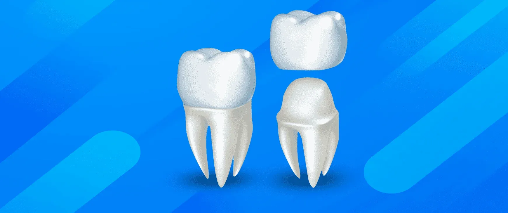
For teeth that have suffered moderate to severe structural damage but have a healthy or successfully treated root, full-coverage dental crowns are the ultimate mechanical savior. A crown acts exactly like a custom-made protective helmet, binding the cracked segments tightly together and preventing the split from widening under the massive, repetitive forces of chewing. High-grade zirconia and porcelain crowns offer unmatched durability while perfectly mimicking the natural translucency of human enamel.
If the damage is purely cosmetic and strictly limited to the front teeth—such as a chipped incisor from a minor fall—porcelain veneers present a highly conservative and deeply aesthetic solution. Veneers are ultra-thin ceramic shells permanently bonded to the front surface of the tooth, hiding the chip while requiring minimal removal of healthy tooth structure. Both of these advanced options are frequently utilized in comprehensive smile design protocols to restore complete facial harmony.
Extraction and Implants: When a Tooth Fracture Is Unsavable
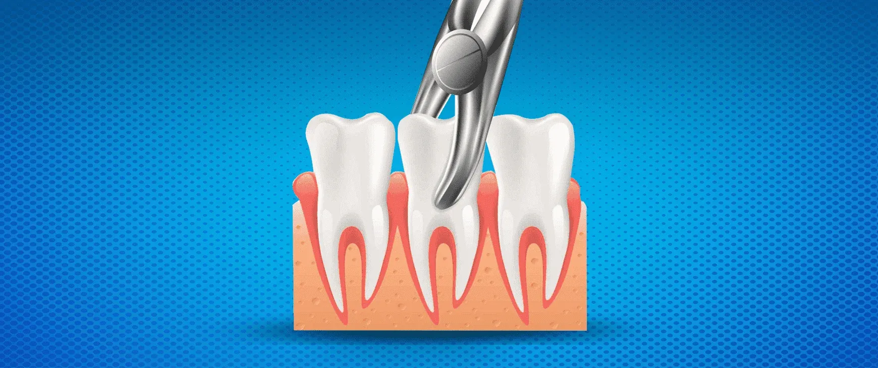
Unfortunately, despite the best efforts of modern medicine, dentists cannot save every broken tooth. If a vertical crack extends deep below the gum line straight into the jawbone, or if the tooth is split completely in half, extraction is the only medically sound and responsible choice. Leaving a split root from a vertical tooth fracture in the jaw guarantees a massive, bone-destroying bacterial abscess that will eventually spread to neighboring tissues.
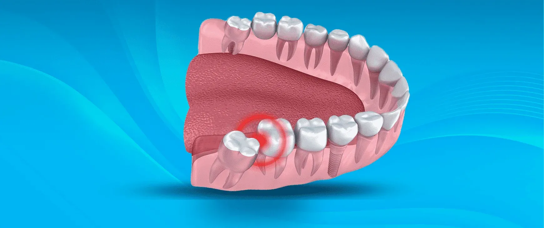
Once the unsavable root is gently and atraumatically extracted, the gold standard for replacement is an artificial titanium root. Placing an implant immediately preserves the jawbone volume and actively prevents adjacent teeth from drifting into the empty socket. After the titanium firmly integrates with the bone, a permanent ceramic crown is attached, fully restoring the patient's natural biting force and confident aesthetic appearance.
Can You Prevent Future Dental Structural Failures?

While genuine accidents certainly happen, the vast majority of mechanical dental failures are entirely preventable with conscious, daily lifestyle adjustments. The most effective preventative measure is abandoning bad oral habits that subject your enamel to unnatural, highly destructive shear forces. Opening hard plastic packaging with your teeth, chewing on metal pens, or crunching down on ice cubes are guaranteed ways to invite a structural disaster.
Regular, bi-annual dental checkups represent your absolute best line of defense. During these routine visits, your dentist can accurately identify failing old silver fillings or microscopic craze lines before they rapidly propagate into full-blown vertical splits. Replacing a massive, leaking filling with a protective crown before the tooth physically breaks is the ultimate and most cost-effective form of preventive dentistry.
The Impact of Bruxism on Long-Term Tooth Fracture Risks
Bruxism, the subconscious grinding and violent clenching of teeth during sleep, is a massive global epidemic driven primarily by modern psychological stress. Human jaws can exert hundreds of pounds of pressure, and when this devastating force is unleashed on enamel night after night, the structural fatigue is undeniable. The constant mechanical friction wears away the protective enamel, leaving the weaker internal dentin exposed and highly vulnerable to shattering.
Patients who suffer from untreated bruxism inevitably develop deep horizontal micro-cracks at the gum line and severely flattened chewing surfaces over the years. To definitively halt this mechanical destruction and minimize tooth fracture risks, a custom-fitted acrylic night guard is absolutely essential. This simple appliance acts as a shock absorber, distributing the destructive bite forces evenly and preventing teeth from grinding directly against one another.
Aesthetic Recovery Following Anterior Dental Trauma
Experiencing a visible break on a highly prominent front tooth is deeply traumatic and instantly impacts a patient's social and professional confidence. Fortunately, anterior cosmetic restorations have advanced to a point where the medical repair is optically indistinguishable from natural, unbroken enamel. Dentists use advanced, light-curing composite resins that are perfectly color-matched and sculpted directly onto the tooth to fix minor chips in a single, painless visit.
For much more extensive anterior damage, laboratory-milled ceramics are strictly required to flawlessly recreate the complex anatomy of front teeth. The ultimate clinical goal is not just to close the broken gap, but to seamlessly match the surface texture, light reflection, and exact color shade of the adjacent healthy teeth. This blend of artistic and clinical precision ensures the patient can smile broadly again without any hesitation.
Why Traveling Abroad for Tooth Fracture Repair Is Popular
The heavy financial burden of complex dental emergencies, especially those requiring multiple root canals, surgical extractions, crowns, or implants, can be overwhelmingly high in many Western countries. Standard insurance policies frequently cap out rapidly or outright refuse to cover major restorative work, leaving traumatized patients with massive out-of-pocket expenses. This harsh economic reality has driven a massive surge in medical tourism for immediate dental trauma management.
Thousands of patients are now actively researching the precise logistics of getting teeth done in turkey to access premium, high-tech emergency care at a fraction of the domestic cost. By meticulously selecting the best dental clinic in turkey, international patients ensure they receive immediate, world-class surgical and restorative treatments without the devastating financial strain, all while recovering in a modern, hospitality-focused clinical environment.
Pediatric Dental Injuries: Managing Broken Teeth in Children
Children are incredibly prone to facial trauma due to constant falls, sports, and highly active play. When a child experiences a break, the clinical approach differs drastically from adults, primarily because primary (baby) teeth have proportionally very large central nerve chambers. A seemingly minor superficial chip on a baby tooth can easily expose the pulp, leading to rapid bacterial infection and severe, unrelenting pain for the child.
If a permanent tooth is injured in a young child, preserving the vitality of the internal nerve is the absolute highest medical priority. Because the tooth root is still actively growing and closing within the jawbone, an early root canal can permanently halt jaw development in that specific area. Pediatric dentists use specialized pulp-capping medicaments to heal the damaged nerve and encourage the root to finish its natural biological maturation process.
Key Steps for Parents During Dental Trauma
Remaining calm is the absolute first duty of a parent, as children will instantly mirror adult panic and hysteria. Immediately assess the mouth for active bleeding and apply gentle pressure with clean gauze. Quickly locate any broken enamel fragments, place them in milk, and proceed directly to an emergency pediatric dentist. Time is of the essence when attempting to save the vitality of a young, traumatized nerve.
Post-Treatment Care for a Restored Tooth Fracture
Walking out of the clinic with a beautifully restored tooth is only the very beginning; protecting that significant medical investment requires strict adherence to daily home care protocols. In the days following a major tooth fracture repair or crown placement, the surrounding gum tissue will naturally be highly inflamed. Patients must stick strictly to a soft diet and completely avoid chewing directly on the newly restored area until the periodontal ligaments fully settle and the inflammation subsides.
Long-term maintenance is largely identical to caring for natural teeth but requires a much heightened daily awareness. Flossing daily is strictly non-negotiable, as plaque accumulation at the margin of a new crown will rapidly cause secondary decay beneath the restoration. Wearing your prescribed night guard and attending professional cleanings every six months will ensure your newly fortified tooth remains entirely intact and infection-free for decades.
- Maintain strict flossing habits around the exact margins of new crowns.
- Avoid intensely hard or highly sticky foods that could dislodge temporary or permanent restorations.
- Wear a custom night guard religiously if you are diagnosed with nocturnal bruxism.
- Attend biannual professional cleanings and X-rays to medically monitor the restoration's ongoing integrity.
Doctor Profile

This content has been evaluated in accordance with Hospitadent Medical Publication Board standards for medical accuracy, currency and patient information.
Our Awards
See All ›2019 – 2020
Health Tourism Champion
Since its founding, Hospitadent has provided oral and dental care to patients travelling from every continent for health tourism. In the Service Exporters' Association research of 2019 and 2020, we ranked first in the Dental Health Services category. Within the same report we placed 15th among all healthcare institutions in 2019 and moved up to 10th in 2020. With uncompromising customer satisfaction, Hospitadent remains the healthcare institution that treats the highest number of international oral and dental patients.
2013
TEMOS International Quality Certificate
Guided by dedicated international departments, Hospitadent welcomes patients from abroad without any language barriers, offering assistance with accommodation, city and Bosphorus tours, concierge guidance, treatment logistics and medication support to keep satisfaction at its peak. With the goal of carrying the same quality standards to both Turkish and foreign patients, we received the TEMOS "International Quality in Dental Care" certificate in 2013—an accreditation granted by the organization that evaluates and certifies worldwide patient service quality based on the "Made in Germany" quality seal.
2020
Turquality Brand Support Program
For 16 years, the Turquality Brand Support Program has helped Turkish products build and promote their image abroad. Dental Group Hospitadent earned the right to join this initiative, taking an important step on its branding journey. Being the first dental healthcare group in a program that includes only five hospitals from the healthcare sector proves not only the quality image we project, but also the high standards we uphold.
2018
Stevie Awards – Most Innovative Customer Service
The Stevie Awards, recognized worldwide for honoring outstanding organizations and leaders, is the only international business awards program. In 2018, Hospitadent became the first hospital in Turkey to win a Stevie Award by receiving the "Most Innovative Customer Service of the Year" prize. Signature experiences such as Smiling Center, Digital Smile Design and Dental SPA played a decisive role in earning this accolade.
2019 – 2020
Service Exporters Association Award
Serving dental tourists from all over the world since its foundation, Hospitadent was honored by the Service Exporters' Association in the Champions of Service Exports program, which celebrates the rising value of Turkey's service industries. Thanks to its contribution to the national economy, Hospitadent received a certificate of achievement and ranked 15th among general healthcare institutions in 2019 and 10th in 2020, continuing to lead oral and dental treatments with an unwavering focus on patient satisfaction.
2021
Great Place To Work® Certified
Based on the employee experience and satisfaction survey completed by our teams, GPTW Turkey evaluated Hospitadent and awarded us the "Great Place To Work® Certified" distinction. As the first and only dental hospital holding this leading workplace culture certificate, Hospitadent continues its journey toward many more achievements.